NUHCS and NUH launch novel Photon-Counting CT to advance high-risk cardiac care
Published on 22 May 2026
The National University Heart Centre, Singapore and National University Hospital introduced the new photon-counting imaging technology on 18 May, to drive non-invasive diagnostics and collaborative innovation in cardiac care.
The Photon-Counting CT delivers unprecedented anatomical clarity, reducing invasive angiograms to safely protect high-risk cardiac patients.
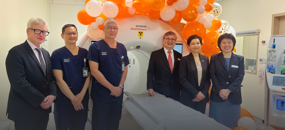
SINGAPORE - As Singapore faces the demographic reality of a rapidly ageing population, the systemic burden of complex cardiovascular disease continues to escalate. In response, the National University Heart Centre, Singapore (NUHCS) and National University Hospital (NUH) recently launched its first Photon-Counting Computed Tomography (PCCT) scanner.
NUHCS is the first tertiary centre in Singapore to deploy the PCCT capability for a high workload of complex cardiac cases.
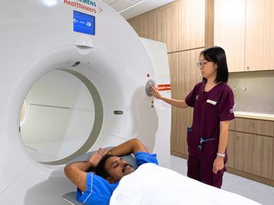
Equipped with advanced photon-counting detector technology, the new dual-source CT scanner fundamentally shifts how clinicians at NUHCS diagnose and manage cardiovascular diseases for high-risk patients. It offers unprecedented image clarity while significantly reducing the need for invasive diagnostic procedures.
Traditionally, heavily calcified vessels or existing stents have made it difficult to obtain clear diagnostic images. This visual barrier often means high-risk patients may have to undergo invasive catheter angiograms for cardiologists to visualise their coronary anatomy before treatment.
"This is a paradigm shift in the way scanners are designed," explained A/Prof James Yip, Executive Director and Senior Consultant, Department of Cardiology, NUHCS. "Now we can be closer to 95 per cent sure that a patient does not need an angiogram. It reduces the need for an unnecessary procedure that has risks, offering higher safety and greater accuracy for our most fragile patients."
A paradigm shift in precision and safety
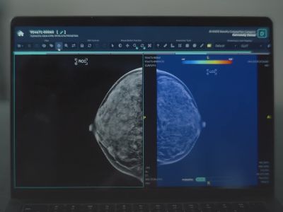
To overcome these visual barriers, the PCCT system departs from how traditional CT scanners work. Instead of measuring X-ray energy the way a bucket measures rain — collecting everything together and reading the total volume — the PCCT counts each individual X-ray particle one by one and measures its energy.
"This technology represents a significant advancement in how we image our patients and how we practise radiology," shared Prof Khong Pek Lan, Head and Senior Consultant, Department of Diagnostic Imaging, NUH. "It allows us to visualise structures in exceptionally high resolution, down to 0.2mm, and allows multi-spectral imaging, which provides accurate characterisation of tissue and materials."
This molecular-level precision translates directly into patient safety. Every CT scan requiring contrast dye carries risks related to radiation exposure and the dye itself, which in high doses may potentially harm the kidneys. The new PCCT system features a multi-use precision contrast injector to tailor exactly how much dye and radiation each patient receives.
"With Photon-Counting CT, we can find disease earlier, diagnose more confidently, and do so more safely than ever before," noted Adj A/Prof Lynette Teo, Senior Consultant, Department of Diagnostic Imaging, NUH.
"The scanner is powerful, but we need to use this technology responsibly," said Adj A/Prof Teo. "This new scanner will specifically benefit our vulnerable populations and high-risk patients, providing datasets that are two to three times larger and clearer than our original scanners."
Bringing technology to life for vulnerable patients
The tangible impact of this technology was recently demonstrated during a Transcatheter Aortic Valve Implantation (TAVI) planning scan. The patient was a frail 90-year-old gentleman suffering from severe aortic stenosis, hearing impairment, and underlying kidney issues.
His narrowed heart valve resulted in a dangerously low cardiac output. Consequently, he could not safely receive the standard preparatory medications typically required to slow the heart rate before a conventional CT scan.
Using the PCCT, the multidisciplinary care team obtained outstanding images of his coronary arteries, aortic valve, and aorta in a single scan without any preparatory medications. The scan required just 55mls of contrast dye and a minimal radiation dose of 10mSv, equivalent to approximately 500 standard chest X-rays.
"We managed to do this scan for him with a very low amount of contrast dye, without having to slow down his heart rate," highlighted A/Prof Yip. "He was a very fragile patient, but he did not suffer any kidney problems after the scan."
Following the non-invasive scan, the patient successfully underwent his TAVI procedure and recovered well, requiring only a short two-day hospital stay.
Advancing academic medicine and future-ready care
The ability to rely safely on CT imaging for complex interventions represents a major shift in clinical workflows. The PCCT scanner not only enhances patient care but also serves as a vital platform for resident training and clinical research in dose optimisation.
As Singapore’s healthcare system adapts to the demands of an ageing society, these new advances translate into a safer, smoother, and more efficient care experience. Ultimately, these developments underscore NUHCS and NUH’s commitment to delivering the best in cardiovascular care, and to continually build upon its capabilities to meet Singapore’s evolving healthcare needs.
To download the PDF version of the media release, click here.